Laboratory Manual for Anatomy & Physiology: Article Plan
Pearson’s new 10th edition (1292026375, 9781292026374) offers extensive instructor support, aiding quizzing. Seeley’s 12th edition provides kidney anatomy exercises;
Anatomy & Physiology laboratory manuals are crucial companions to lecture courses, bridging theoretical knowledge with practical application. These manuals, like Pearson’s 10th edition and Seeley’s, guide students through dissections, microscopy, and physiological experiments. They often include pre-lab quizzes and assessments to reinforce learning. The University of Georgia utilizes a transformed, open-source manual.
Instructors benefit from accompanying guides offering answer keys and time schedules. These resources enhance teaching and ensure effective lab sessions, fostering a deeper understanding of the human body.
II. Importance of Lab Work in A&P
A&P lab work transcends textbook learning, providing hands-on experience with anatomical structures and physiological processes. Dissection, utilizing manuals with detailed guidelines – like those for laboratory animal dissection – allows for spatial understanding. Microscopy, a core skill, is honed through techniques like wet mounts and staining.
These experiences solidify concepts, develop critical thinking, and prepare students for clinical applications, making the laboratory an indispensable component of A&P education.
III. Core Components of a Typical A&P Lab Manual
A&P lab manuals commonly feature safety regulations and guidelines, prioritizing a secure learning environment. Pre-lab quizzes and assessments, often with instructor’s guides providing answers, gauge preparedness and reinforce concepts. Content includes exercises on cells, tissues, skeletal, muscular, and nervous systems.
These manuals often cover broad topics, suitable for both one and two-semester courses, facilitating comprehensive anatomical and physiological exploration.
A. Safety Regulations & Guidelines
A&P lab manuals prioritize safety, outlining crucial regulations for handling specimens, chemicals, and equipment. Guidelines emphasize proper dissection techniques, minimizing risks during animal studies – a feature found in some editions.
Protocols for microscope usage, including slide handling and staining procedures, are detailed. Emphasis is placed on personal protective equipment (PPE) and waste disposal, ensuring a secure and responsible laboratory experience for all students.
B. Pre-Lab Quizzes & Assessments
A&P lab manuals increasingly incorporate pre-lab quizzes to gauge student preparedness and reinforce foundational knowledge. These assessments, often available with instructor support materials – like those in Pearson’s editions – test understanding of procedures and concepts before practical work begins.
Post-lab assessments evaluate comprehension and skill application. Instructor’s guides, such as for the 5th edition, provide answer keys and time estimations for these evaluations, streamlining the teaching process.

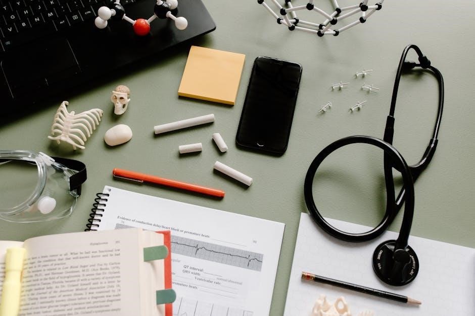
IV. The Microscope: A Foundational Tool
A&P lab manuals dedicate significant space to microscopy, recognizing its central role in anatomical study. Chapter 2 in many manuals, including Pearson’s, focuses on the microscope’s parts and functions, essential for observing tissues and cells.
Labs detail techniques like wet mount preparation and staining, crucial for visualization. Mastering microscopy is foundational, enabling students to analyze histological slides and understand cellular structures effectively.
A. Parts of a Microscope & Their Functions
Laboratory manuals meticulously detail each microscope component. Pearson’s and Seeley’s editions explain the function of the ocular lens, objective lenses, stage, and condenser. Students learn how adjusting these parts impacts magnification and resolution.
Understanding the diaphragm’s role in light control and the coarse/fine focus knobs for clarity is emphasized. Manuals often include diagrams labeling each part, reinforcing anatomical knowledge of this vital tool.
B. Microscopy Techniques (Wet Mounts, Staining)
Anatomy & Physiology laboratory manuals guide students through essential microscopy techniques. Pearson’s and similar texts detail creating wet mounts for observing living cells, emphasizing proper slide preparation.
Crucially, manuals explain various staining procedures – their purpose, application, and impact on cellular visibility. Students learn how stains enhance contrast, revealing intricate cellular structures. These techniques are foundational for histological analysis and understanding tissue characteristics.
V. Histology: The Study of Tissues
Laboratory manuals for Anatomy & Physiology dedicate significant sections to histology, the microscopic study of tissues. These resources guide students in identifying the four primary tissue types: epithelial, connective, muscle, and nervous.
Manuals provide detailed descriptions and visual aids for recognizing each tissue’s unique characteristics. Students learn to differentiate between subtypes, like various epithelial arrangements, and understand how tissue structure relates to function. Practical exercises reinforce microscopic identification skills.
A. Types of Epithelial Tissue & Identification
Anatomy & Physiology laboratory manuals meticulously cover epithelial tissues, categorized by cell shape (squamous, cuboidal, columnar) and layering (simple, stratified, pseudostratified). Students learn to identify these types under a microscope, noting key features like nuclei position and cell junctions.
Manuals often include illustrative diagrams and photomicrographs, aiding in accurate recognition. Exercises focus on distinguishing between different epithelial subtypes and correlating their structure with specific functions within the body.
B. Connective Tissue Classification & Characteristics
Laboratory manuals detail connective tissue classification – including connective tissue proper (loose, dense), supporting connective tissues (cartilage, bone), and fluid connective tissues (blood). Students learn to identify these tissues based on their cellularity, matrix composition, and fiber types (collagen, elastic, reticular).
Practical exercises involve microscopic observation, comparing and contrasting tissue structures. Manuals emphasize the relationship between connective tissue characteristics and their specific roles in support, protection, and transport.
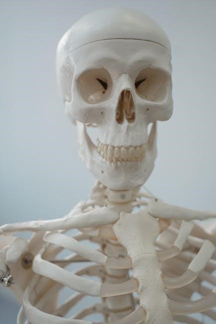
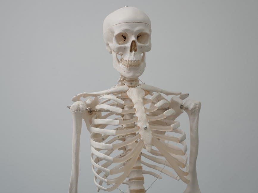
VI. Skeletal System Laboratory Exercises
Anatomy & Physiology lab manuals guide students through bone identification, focusing on anatomical landmarks and features. Exercises involve assembling skeletal models, articulating bones to demonstrate joint types, and classifying articulations based on structure and function.
Students learn to differentiate between various bone shapes and relate them to their specific roles. Practical applications include analyzing skeletal trauma and understanding bone pathologies, enhancing clinical relevance.
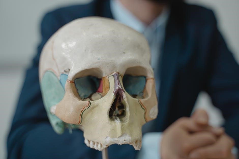
A. Bone Identification & Anatomy
Laboratory exercises emphasize identifying individual bones – cranial, vertebral column, appendicular skeleton – using models and diagrams. Students learn anatomical landmarks: processes, foramina, tubercles, and fossae, relating structure to function.
Manuals often include detailed illustrations and labeling exercises. Understanding bone classifications – long, short, flat, irregular – is crucial. Practical application involves recognizing bones in radiographs and relating anatomical features to clinical scenarios.
B. Articulations & Joint Classification
Lab sessions focus on classifying joints structurally – fibrous, cartilaginous, synovial – and functionally – synarthrotic, amphiarthrotic, diarthrotic. Students examine joint models, identifying key features like articular cartilage, joint capsules, and ligaments.
Manuals detail specific joint types (e.g., hinge, ball-and-socket) and their range of motion. Practical exercises involve palpating joints and analyzing movements. Understanding joint stability and common injuries is emphasized.
VII. Muscular System Dissection & Study
Laboratory exercises involve identifying major skeletal muscles, tracing their origins and insertions, and analyzing their actions. Dissection (when applicable) provides hands-on experience with muscle structure and relationships.
Manuals guide students through microscopic examination of muscle tissue types – skeletal, smooth, cardiac – noting key characteristics. Emphasis is placed on understanding muscle fiber arrangement and its impact on force production. Clinical correlations, like muscle strains, are often included.
A. Muscle Tissue Types & Microscopic Structure
Laboratory manuals facilitate the microscopic identification of skeletal, smooth, and cardiac muscle tissues. Students learn to distinguish features like striations (present in skeletal and cardiac), nuclei arrangement, and cell shape.
Detailed diagrams and photomicrographs aid in recognizing key structures within each tissue type. Emphasis is placed on correlating microscopic anatomy with functional properties. Exercises often involve comparing and contrasting the three muscle types, noting their unique adaptations.
B. Major Muscle Identification & Function
Laboratory exercises focus on identifying major skeletal muscles on anatomical models and, when possible, through dissection. Students learn origins, insertions, and actions of key muscles like biceps brachii, rectus femoris, and gastrocnemius.
Manuals often include tables summarizing muscle characteristics and functional groups. Activities may involve tracing muscle actions on diagrams or performing simple movements to observe muscle contractions. Understanding the relationship between muscle structure and function is paramount.
VIII. Nervous System Exploration
Laboratory sessions delve into the intricate world of the nervous system, beginning with brain dissections to identify anatomical structures like the cerebrum, cerebellum, and brainstem. Students trace pathways and correlate structure with function.
Manuals guide identification of spinal cord segments and associated nerves. Activities may include reflex arc experiments and sensory perception testing. Emphasis is placed on understanding the organization and communication within the nervous system, crucial for physiological processes.
A. Brain Dissection & Anatomical Structures
Brain dissection is a cornerstone of nervous system labs, allowing students to directly visualize anatomical structures. Laboratory manuals guide identification of key regions – the cerebrum, cerebellum, brainstem, and associated lobes.
Students learn to differentiate gray and white matter, locate major sulci and gyri, and identify cranial nerves. Emphasis is placed on correlating structural features with their functional roles in sensation, movement, and cognition. Careful dissection and precise labeling are essential.
B. Spinal Cord & Nerve Identification
Laboratory exercises focus on identifying the external and internal structures of the spinal cord. Students learn to distinguish between the dorsal and ventral horns, and locate the central canal. Manuals guide the tracing of nerve roots and their exit points from the spinal column.
Identification of major peripheral nerves – such as the sciatic, ulnar, and median nerves – is crucial. Emphasis is placed on understanding the relationship between nerve structure and its innervation territory. Careful observation and accurate labeling are paramount.
IX. Cardiovascular System Investigations
Lab sessions involve detailed heart dissection, revealing chambers, valves, and major vessels. Students trace blood flow pathways and identify coronary arteries. Manuals guide analysis of blood samples, determining components like erythrocytes, leukocytes, and platelets.
Investigations include observing blood vessel anatomy – arteries, veins, and capillaries – and correlating structure with function. Emphasis is placed on understanding the cardiac conduction system. Accurate observation and proper handling of specimens are essential for learning.
A. Heart Dissection & Blood Vessel Anatomy
Laboratory manuals provide step-by-step guidance for heart dissection, identifying chambers, valves (tricuspid, mitral, aortic, pulmonary), and major vessels like the aorta and vena cava. Students carefully trace the path of blood flow through the heart.
Detailed study of blood vessel anatomy includes differentiating arteries, veins, and capillaries based on structural features. Emphasis is placed on correlating anatomical structure with physiological function, understanding how vessel design supports blood circulation. Proper technique ensures accurate observation.
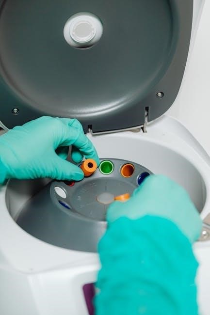
B. Blood Analysis & Component Identification
Laboratory exercises focus on blood analysis, utilizing techniques like hematocrit determination and blood typing. Students learn to identify blood components – erythrocytes, leukocytes, and thrombocytes – under a microscope, noting their distinct characteristics.
Manuals guide identification of different leukocyte types (neutrophils, lymphocytes, monocytes, eosinophils, basophils) and their roles in immunity. Analysis includes observing blood smears and interpreting results, connecting structure to function. Accurate identification is crucial for understanding blood’s role in health.
X. Respiratory System Analysis
Laboratory manuals detail lung dissection procedures, enabling students to visualize anatomical structures like bronchi, bronchioles, and alveoli. Exercises involve tracing the respiratory pathway – nasal cavity to alveoli – and identifying key muscles involved in breathing.
Students analyze lung capacity using spirometers, correlating results with anatomical features. Manuals often include microscopic examination of lung tissues, identifying alveolar structure. Understanding the relationship between structure and function is emphasized, linking anatomy to respiratory physiology.
A. Lung Dissection & Respiratory Pathway Tracing
Laboratory manuals guide students through careful lung dissection, identifying major structures like the trachea, bronchi, and pulmonary vessels. Emphasis is placed on observing the branching pattern of airways and the delicate alveolar structure. Tracing the respiratory pathway – from nasal cavity through the pharynx, larynx, and lungs – reinforces anatomical understanding.
Students learn to identify muscles involved in respiration, like the diaphragm and intercostals. Detailed illustrations and diagrams within the manual aid in accurate identification and pathway visualization.
XI. Digestive System Examination
Anatomy and physiology laboratory manuals facilitate a comprehensive examination of the digestive system through organ dissection. Students meticulously dissect organs – stomach, small and large intestines, liver, pancreas – observing anatomical relationships. Emphasis is placed on identifying structures like villi, papillae, and sphincters.
The manuals guide tracing the alimentary canal, understanding peristalsis, and identifying accessory organs’ roles. Detailed diagrams and instructions ensure accurate dissection and anatomical comprehension.
A. Organ Dissection & Anatomical Relationships
Laboratory manuals guide students through detailed organ dissection of the digestive system – stomach, intestines, liver, and pancreas; Emphasis is placed on identifying anatomical structures like villi, papillae, and sphincters, fostering a deeper understanding. Students trace the alimentary canal, observing peristalsis and accessory organ roles.
Manuals provide clear instructions and diagrams, ensuring accurate dissection and comprehension of anatomical relationships within the digestive tract. This hands-on approach reinforces theoretical knowledge.
XII. Urinary System Studies
Laboratory exercises focus on kidney anatomy, meticulously examining the renal cortex, medulla, and pelvis. Students identify nephrons – glomeruli, tubules – and trace urine formation pathways. Manuals guide dissection, revealing ureters, bladder, and urethra structures.
Activities include microscopic observation of kidney tissues and analysis of urine composition. Seeley’s lab manual provides detailed kidney anatomy illustrations, enhancing comprehension of this vital system’s function.
A. Kidney Anatomy & Nephron Structure
Labs emphasize identifying kidney regions: cortex, medulla, renal pelvis. Students dissect kidneys, tracing blood flow through afferent/efferent arterioles and glomeruli. Microscopic studies reveal nephron components – Bowman’s capsule, proximal/distal convoluted tubules, loop of Henle, and collecting ducts.
Manuals, like Seeley’s, provide detailed diagrams illustrating nephron structure and function. Exercises focus on correlating anatomy with urine formation processes, enhancing understanding of renal physiology.
XIII. Reproductive System Investigations
Laboratory exercises involve identifying male and female reproductive organs through models and diagrams. Students examine testes, ovaries, uterus, and associated structures, learning their anatomical relationships. Histological slides reveal gamete production sites – seminiferous tubules and ovarian follicles.
Manuals guide dissection (if applicable) and microscopic observation, correlating structure with hormonal control and reproductive functions. Emphasis is placed on understanding the physiological basis of reproduction.
A. Male & Female Reproductive Organ Identification
Lab manuals facilitate identifying key male structures: testes, epididymis, vas deferens, seminal vesicles, prostate, and penis. Female organs include ovaries, fallopian tubes, uterus, cervix, and vagina. Students utilize anatomical models, charts, and potentially preserved specimens.
Exercises focus on locating each organ, noting its shape, size, and connections. Understanding the pathway of sperm and oocytes is crucial. Detailed diagrams aid in visualization and accurate identification.

XIV. Sensory System Experiments
Laboratory exercises explore sensory perception and reflexes, vital components of physiology. Students test visual acuity, auditory thresholds, and tactile discrimination using standardized procedures; Experiments assess taste and smell identification, exploring receptor distribution.
Reflex arc investigations, like the patellar reflex, demonstrate neural pathways. Manuals guide measuring reaction times and analyzing sensory adaptation. Data analysis helps correlate structure with function, enhancing understanding of the nervous system’s role.
A. Testing Sensory Perception & Reflexes
Experiments meticulously test visual acuity, auditory thresholds, and tactile discrimination, employing standardized protocols. Students identify taste and smell, mapping receptor distributions across the tongue and nasal cavity. Lab manuals detail procedures for assessing two-point discrimination and pinpointing blind spots.
Reflex arc investigations, notably the patellar reflex, illustrate neural pathways. Reaction time measurements and sensory adaptation analyses provide quantitative data, linking structure to function.
XV. Utilizing Virtual Labs & Digital Resources
Modern A&P lab manuals increasingly integrate virtual simulations, offering alternatives to traditional dissection. Digital resources, like interactive anatomy models, enhance visualization and spatial understanding. Online quizzes and assessments provide immediate feedback, reinforcing learning.
Platforms offer pre-lab tutorials and post-lab review materials. Instructors leverage these tools for remote learning and to supplement hands-on experiences, improving student engagement and accessibility.
XVI. Dissection Techniques & Best Practices
Effective dissection requires careful planning and adherence to safety protocols. Manuals often provide detailed guidelines, mirroring those found in physiology laboratory manuals for animal dissection. Proper tool handling, precise incisions, and respectful treatment of specimens are crucial.
Students should learn anatomical relationships systematically, documenting observations meticulously. Instructors emphasize minimizing tissue damage and maintaining a clean, organized workspace for optimal learning outcomes.
XVII. Common Lab Errors & Troubleshooting
Frequent errors in A&P labs include misidentification of structures, improper microscope usage, and inaccurate measurements. Troubleshooting often involves reviewing procedures, seeking instructor guidance, and verifying observations with peers.
Manuals may include sections addressing common pitfalls, like staining inconsistencies or dissection inaccuracies. Students should learn to recognize and correct these errors, fostering critical thinking and problem-solving skills essential for success in the course.
XVIII. Instructor Resources & Support Materials
Comprehensive instructor resources are vital for effective A&P lab teaching. Pearson manuals, like the 10th edition, boast extensive support for easier quizzing and assessment creation. Instructors benefit from pre-lab answer keys, estimated time schedules, and detailed dissection guidelines.
Additional materials may include image banks, lecture outlines, and online resources to enhance teaching and student learning. These support systems streamline lab preparation and ensure a consistent, high-quality educational experience.
XIX. Current Editions & Popular Manuals (Pearson, Seeley’s)
Pearson currently offers the 10th edition of its A&P lab manual (1292026375, 9781292026374), known for its comprehensive coverage and instructor support. Seeley’s Anatomy and Physiology Lab Manual is available in its 12th edition, providing detailed exercises, including those focused on kidney anatomy.
Both manuals are widely adopted in university courses, offering students practical experience with dissections and microscopic analysis; These editions frequently update content to reflect current scientific understanding.
XX. Future Trends in A&P Lab Manuals
The integration of virtual labs and digital resources is a prominent trend, supplementing traditional dissection and microscopy. Expect increased use of augmented and virtual reality to enhance learning and accessibility. Manuals will likely feature more interactive assessments and personalized learning pathways.
Focus will shift towards clinical correlations and real-world applications, bridging the gap between theory and practice. Open Educational Resources (OER) are also gaining traction, offering cost-effective alternatives.